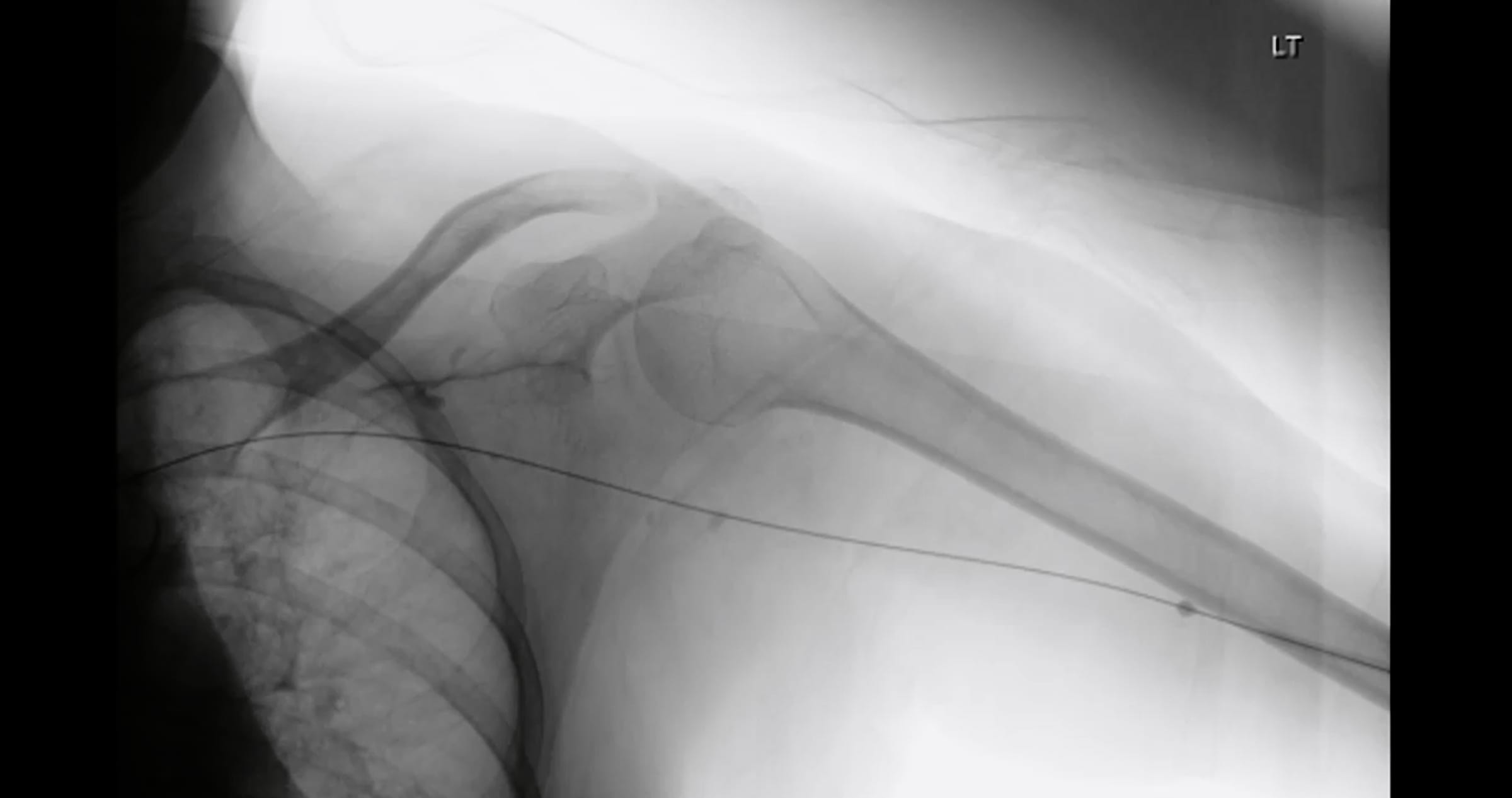
A graftogram is a fluoroscopic imaging study in which a vascular specialist injects iodinated contrast dye through a needle placed in an arteriovenous graft.
The images follow the dye from the arterial anastomosis, through the synthetic conduit, and out to the central veins. Stenosis, thrombus, and pseudoaneurysm all become visible.
Our physicians perform graftograms at Soris Pain & Vascular Institute. Patients with a native arteriovenous fistula receive a Fistulogram instead.
A graft is a synthetic tube sewn between an artery and a vein. It never matures into living vessel wall, so the vein where it drains reacts to the flow by thickening.
That venous anastomosis is where most graft stenoses form. Soris Pain & Vascular Institute images it directly, because a graft that narrows there can clot within days.
Soris Pain & Vascular Institute combines diagnostic imaging with same-session treatment. When the graftogram shows a narrowing, the team performs balloon Angioplasty immediately rather than booking a second visit.